Aggressive solid tumors such as prostate cancer commonly develop an acidic microenvironment with pH 6.5–7.2 due to their heterogeneous perfusion, high metabolic activity, and rapid cell proliferation. Our team has developed an innovative methodology and protocol for enhanced detection of aggressive prostate cancer by utilizing hyperpolarized (HP) 13C-bicarbonate for clinical MRI. This method relies on the rapid breakdown of hyperpolarized 1,2-glycerol carbonate (carbonyl-13C) via base-catalyzed hydrolysis to produce HP 13C-carbonate, which is neutralized and converted to HP 13C-bicarbonate. After administration, HP 13C-bicarbonate equilibrates with HP 13CO2 and enables the imaging of the extracellular pH of prostate cancer tumors. We have validated the method’s reliability and accuracy in measuring the acidic microenvironment of healthy kidney and prostate tumor tissues. Our method has been FDA approved as an investigational new drug and currently in ongoing clinical trial (NCT05851365).
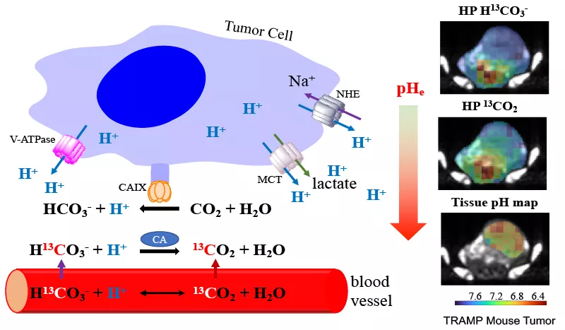
Figure: Hyperpolarized (HP) 13C extracellular pH (pHe) MRI imaging. Left: Extracellular acidification of the tumor microenvironment and the mechanism of imaging interstitial pH using hyperpolarized H13CO3–. Note: NHE, Na+/H+exchanger; MCT, monocarboxylate transporter; CAIX, carbonic anhydrase IX; LDH, lactate dehydrogenase; and V-ATPase, vacuolar H+-ATPase. Right: The figures on the top, middle, and bottom show the 13C MRI images of HP 13C-bicarbonate, HP 13CO2, and pHe map overlaid with anatomical proton MRI figure of a later stage tumor of the transgenic adenocarcinoma of the mouse prostate (TRAMP) model, respectively.
We are actively recruiting for patients. For more information on the clinical trial, please visit ClinicalTrials.gov.
For more information, refer to our published work:
- Mu C, Liu X, Riselli A, Slater J, Escobar E, Dang D, Drapeau S, Delos Santos R, Andosca S, Nguyen H, Larson PEZ, Bok R, Vigneron DB, Kurhanewicz J, Wilson DM, Flavell RR. Protocol for Producing Hyperpolarized 13C-bicarbonate MRI Imaging Contrast for Clinical Imaging of Extracellular pH in Aggressive Tumor. STAR Protocols. https://doi.org/10.1016/j.xpro.2024.103091.
- Mu C, Liu X, Kim Y, Riselli A, Korenchan DE, Bok RA, Delos Santos R, Sriram R, Qin H, Nguyen H, Gordon JW, Slater J, Larson PEZ, Vigneron DB, Kurhanewicz J, Wilson DM, Flavell RR. Clinically Translatable Hyperpolarized 13C Bicarbonate pH Imaging Method for Use in Prostate Cancer. ACS Sens. 2023 Nov 24;8(11):4042-4054. doi: 10.1021/acssensors.3c00851. Epub 2023 Oct 25. PMID: 37878761; PMCID: PMC10683509.
- Mu C, Korenchan DE, Wang S, Wilson DM, Flavell RR. Tumor Microenvironment Biosensors for Hyperpolarized Carbon-13 Magnetic Resonance Spectroscopy. Mol Imaging Biol. 2021 Jun;23(3):323-334. doi: 10.1007/s11307-020-01570-0. Epub 2021 Jan 7. PMID: 33415679; PMCID: PMC8105261.
- Korenchan DE, Flavell RR, Baligand C, Sriram R, Neumann K, Sukumar S, VanBrocklin H, Vigneron DB, Wilson DM, Kurhanewicz J. Dynamic nuclear polarization of biocompatible (13)C-enriched carbonates for in vivo pH imaging. Chem Commun (Camb). 2016 Feb 18;52(14):3030-3. doi: 10.1039/c5cc09724j. PubMed PMID: 26792559; PubMed Central PMCID: PMC4864526. https://pubs.rsc.org/en/content/articlehtml/2016/cc/c5cc09724j
- Korenchan, DE, Gordon, JW, Subramaniam, S, et al. Using bidirectional chemical exchange for improved hyperpolarized [13C]bicarbonate pH imaging. Magn Reson Med. 2019; 00: 1-14. https://doi.org/10.1002/mrm.27780
https://onlinelibrary.wiley.com/doi/full/10.1002/mrm.27780 - Korenchan, D. E., et al. (2019). "Hyperpolarized in vivo pH imaging reveals grade-dependent acidification in prostate cancer." Oncotarget 10(58): 6096-6110. https://www.oncotarget.com/article/27225/
- Korenchan, D. E. and R. R. Flavell (2019). "Spatiotemporal pH Heterogeneity as a Promoter of Cancer Progression and Therapeutic Resistance." Cancers (Basel) 11(7). https://www.mdpi.com/2072-6694/11/7/1026